| Home | E-Submission | Sitemap | Contact Us |  
Journal of Neurocritical Care > Volume 13(2); 2020 > Article
Park, Yoo, Jeong, Lee, Kim, Huh, and Lee: Normal pressure hydrocephalus after gamma knife radiosurgery in a patient with vestibular schwannoma

Abstract

Background

Vestibular schwannoma is a benign, usually slow-growing tumor, which develops from Schwann cells of the eighth cranial nerve.

Case Report

The treatment options for the schwannoma are surgical removal or gamma knife radiosurgery. The treatment of choice depends on the size of the tumor and the level of hearing in the affected ear. After gamma knife radiosurgery, there may be some neurological complications including headache, dizziness, motor or cranial nerve deficits, seizure, carotid artery stenosis, and increased intracranial pressure. Hydrocephalus is a rare complication of gamma knife radiosurgery for vestibular schwannoma.

Conclusion

Here, we report a case of normal pressure hydrocephalus after gamma knife radiosurgery in a patient with vestibular schwannoma.

INTRODUCTION

Vestibular schwannoma (VS) is a benign tumor that grows in Schwann cells of the eighth cranial nerve, and it can be treated by surgical removal or stereotactic radiosurgery including gamma knife surgery. Stereotactic radiosurgery for VS was first attempted in 1969 and has been one of the most active treatments in the last 40 years. The main side effects include facial paralysis, hearing loss, trigeminal neuralgia, and hemifacial spasm [1]. In addition, hydrocephalus is a potential concomitant disease [2-4]. In this article, we report a case of normal pressure hydrocephalus (NPH) in VS that has undergone gamma knife radiosurgery.

CASE REPORT

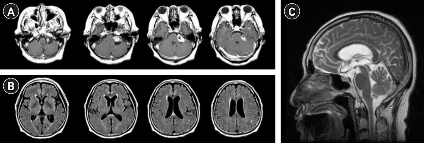
A 67-year-old woman visited the clinic with hearing loss in the left ear. The symptom began 1 month previously. She did not have a specific medical history, except for myasthenia gravis. Vital signs were within normal ranges. Rinne test was normal on both sides. However, the Weber test detected sensorineural hearing loss on the left side. No other abnormalities were observed during other neurological examinations. Magnetic resonance imaging revealed a high signal intensity mass measuring 2.7 cm×3.0 cm with fine enhancement on the left cerebellopontine angle (Fig. 1). This lesion was diagnosed as VS, and gamma knife surgery was performed.
Three months after, the patient visited the clinic again, complaining of progressive cognitive decline, gait disturbance, and urinary incontinence. Neurologic examination revealed normal cranial nerve functions. Motor and sensory functions were intact. Finger-to-nose and heel-to-shin tests were normal. However, she had a magnetic gait with a wide base. Additionally, she kept on losing her balance as she walked. The Timed Up and Go test was measured at 26 seconds. The patient had 2 years of education. Her Korean Mini-Mental Status Examination (K-MMSE) score was 17/30. Moreover, Seoul Neuropsychological Screening Battery (SNSB) showed impaired attention, verbal and visual memory, and executive function (Table 1).
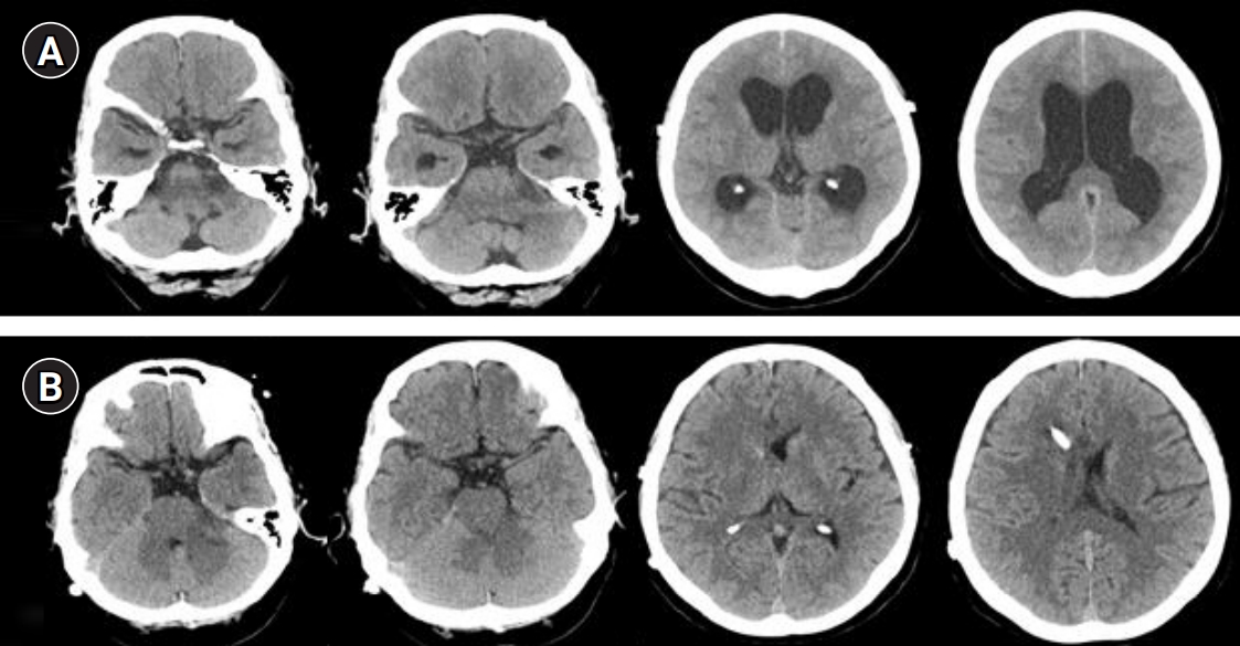
On follow-up computed tomography (CT) scan, the size of the VS decreased from 2.7 cm×3.0 cm to 1.6 cm×2.8 cm, reducing the mass effect to the surrounding brain stem. However, newly developed bilateral ventricular dilatation was observed (Fig. 2). The Evans ratio was 0.37. Cerebrospinal fluid (CSF) drainage was performed, and the pressure was measured at 10 cm H2O. Leukocyte count was 0/mm3, erythrocyte count was 0/mm3, protein concentration was 58 mg/dL, and sugar level was 88 mg/dL (blood sugar, 97 mg/dL), demonstrating mild increases in protein level. After CSF drainage, the patient’s gait and urinary incontinence improved. The Timed Up and Go test was measured again, and the result decreased from 26 to 22 seconds. A ventriculoperitoneal shunt was inserted, resulting in subjective improvements in the gait disturbance. Her follow-up K-MMSE score improved to 22, and visual memory and executive function in the SNSB, performed after 3 months, also improved (Table 1). Another CT scan was conducted, and it revealed that the size of the ventricle had decreased, and the Evans ratio measured 0.21 (Fig. 2).

DISCUSSION

Hydrocephalus has been reported repeatedly in patients with VS who undergo gamma knife radiosurgery [1-5]. A causal relationship, however, has not been identified, and the mechanism remains unclear. The most likely hypothesis for the relationship between gamma knife radiosurgery and hydrocephalus involves tumor necrosis, which causes the protein to deposit in arachnoid granulation and interfere with CSF absorption [3]. In this case, the level of protein was slightly increased, supporting this hypothesis. In addition, fourth ventricle compression [6] or CSF flow alteration in basilar cisterns [7] is considered to be another potential mechanism. However, whether these side effects occur only in radiosurgery, including gamma knife, is still controversial [8,9].
According to a 2012 study [9], 19.9% of patients who underwent gamma knife surgery were diagnosed with hydrocephalus. However, 12.8% were diagnosed before gamma knife surgery and 7.6% occurred after surgery. In the same study, there was a statistically significant risk of the development of hydrocephalus, especially in patients with large tumor masses or who are female. With this in mind, the patient in this case was diagnosed with newly developed NPH, characterized by the traditional triad of gradual memory loss, gait disturbance, and urinary incontinence 3 months after gamma knife surgery. The causal relationship between NPH and gamma knife radiosurgery is further supported by the patient's symptom recovery with the insertion of a ventricular abdominal shunt.
As mentioned above, there have been several reported cases of hydrocephalus developing after radiosurgery for VS. However, in this case, the patient complained of a sudden decline in cognitive function, gait disturbance, and urinary frequency after gamma knife surgery, and the recovery of cognitive function after the shunt operation was confirmed through subjective reports by patients and guardians, as well as objective neuropsychiatric tests. This case is meaningful in emphasizing that neurologists should actively discriminate and exclude secondary correctable causes, such as NPH, when rapid progressive cognitive decline and gait disturbances occur in patients with a history of brain radiosurgery, especially considering its reversibility.

ARTICLE INFORMATION

Ethics statement
The requirements for approval of the Institutional Review Board and informed consent were waived due to the retrospective nature of this study.
Conflict of interest
No potential conflict of interest relevant to this article.
Author contributions
Concepualization: BGY. Project administration: SJ. Visualization: JHL. Writing–origicnal draft: YAP. Writing–review & editing: MKK, SYH, WGL.

Fig. 1.
(A) T1-enhanced images show a mass in the left cerebellopontine angle. (B) Fluid-attenuated inversion recovery images reveal a normal-sized ventricle. (C) T1 midsagittal view does not show any evidence of non-communicating hydrocephalus.
jnc-200014f1.jpg
Fig. 2.
Non-contrast computed tomography (CT) images of the patient. (A) Three months after gamma knife surgery, follow-up CT scans revealed enlarged ventricles. (B) After ventriculoperitoneal shunt insertion, the size of the ventricles normalized.
jnc-200014f2.jpg
Table 1.
Results of the neuropsychological test (SNSB) of pre- and post-ventriculoperitoneal shunt
Variable Preoperative
Postoperative
Raw score %Score Raw score %Score
K-MMSE 17 0.27 23 0.15
Digit span
 Forward 4 26.11 4 26.11
 Backward 0 0.01 2 11.51
K-BNT 24 0.4 25 0.71
RCFT
 Copy 1 0.01 18 10.56
 Immediate recall NA NA 0.5 5.48
 20-Minute-delayed recall NA NA 0 3.92
 Recognition 13 0.82 15 5.94
SVLT
 Immediate recall 11 12.51 12 17.88
 20-Minute-delayed recall 0 0.15 2 3.14
 Recognition 13 0.06 17 9.01
Frontal/executive function
 Contrast program 6 <16 17 <16
 Go-no-go test 3 <16 14 >16
 Fist-edge-palm test Abnormal Abnormal
 Alternating hand movement Abnormal Normal
 Alternating square and triangle Preservation Preservation
 Luria loop Preservation Preservation
COWAT
 Animal 5 0.37 13 33
 Supermarket 3 0.26 9 6.55
 ㄱ 0 1.25 0 1.25
 ㅇ 0 5.59 1 13.35
 ㅅ 0 4.27 0 4.27
 Phonemic total score 0 1.13 1 1.79
K-CWST
 Word reading: number of correct response 45 <16 59 <16
 Word reading: number of error 8 <16 4 <16
 Word reading: response time 120 120
 Color reading: number of correct response 10 0.01 25 0.18
 Color reading: number of error 28 0.01 19 0.01
 Color reading: response time (sec) 120 120
CDR
 Score 1 0.5
 Sum of boxes 5 2
GDS NA 2
B-ADL 13 19

SNSB, Seoul Neuropsychological Screening Battery; K-MMSE, Korean Mini-Mental Status Examination; K-BNT, Korean-Boston Naming Test; RCFT, Rey Complex Figure Test; NA, not applicable; SVLT, Seoul Verbal Learning Test; COWAT, Controlled Oral Word Association Test; K-CWST, Korean-Color Word Stroop Test; CDR, Clinical Dementia Rating; GDS, Global deterioration scale; B-ADL, Basic Activity of Daily Living.

REFERENCES

1. Hasegawa T, Kida Y, Kato T, Iizuka H, Kuramitsu S, Yamamoto T. Long-term safety and efficacy of stereotactic radiosurgery for vestibular schwannomas: evaluation of 440 patients more than 10 years after treatment with Gamma Knife surgery. J Neurosurg 2013;118:557-65.
crossref pmid
2. Cauley KA, Ratkovits B, Braff SP, Linnell G. Communicating hydrocephalus after gamma knife radiosurgery for vestibular schwannoma: an MR imaging study. AJNR Am J Neuroradiol 2009;30:992-4.
crossref pmid pmc
3. Bloch J, Vernet O, Aubé M, Villemure JG. Non-obstructive hydrocephalus associated with intracranial schwannomas: hyperproteinorrhachia as an etiopathological factor? Acta Neurochir (Wien) 2003;145:73-8.
crossref pmid pdf
4. Rogg JM, Ahn SH, Tung GA, Reinert SE, Norén G. Prevalence of hydrocephalus in 157 patients with vestibular schwannoma. Neuroradiology 2005;47:344-51.
crossref pmid pdf
5. Norén G. Long-term complications following gamma knife radiosurgery of vestibular schwannomas. Stereotact Funct Neurosurg 1998;70 Suppl 1:65-73.
crossref pmid
6. Briggs RJ, Shelton C, Kwartler JA, Hitselberger W. Management of hydrocephalus resulting from acoustic neuromas. Otolaryngol Head Neck Surg 1993;109:1020-4.
crossref pmid
7. Steenerson RL, Payne N. Hydrocephalus in the patient with acoustic neuroma. Otolaryngol Head Neck Surg 1992;107:35-9.
crossref pmid
8. Jeon CJ, Kong DS, Nam DH, Lee JI, Park K, Kim JH. Communicating hydrocephalus associated with surgery or radiosurgery for vestibular schwannoma. J Clin Neurosci 2010;17:862-4.
crossref pmid
9. Lee SH, Seol HJ, Kong DS, Nam DH, Park K, Kim JH, et al. Risk factors and tumor response associated with hydrocephalus after gamma knife radiosurgery for vestibular schwannoma. Acta Neurochir (Wien) 2012;154:1679-84.
crossref pmid pdf
Editorial Office
Department of Neurology and Critical Care, Seoul National University Hospital,
101, Daehak-ro, Jongno-gu, Seoul, 03080 Korea
TEL: +82-2-2072-0743 (10:00 AM ~ 4:00 PM) / +82-2-2072-1810   FAX: +82-2-3672-7553    E-mail: office@e-jnc.org

Copyright© Korean Neurocritical Care Society.                Developed in M2PI