INTRODUCTION
Posterior reversible encephalopathy syndrome (PRES) is classically characterized by symmetric vasogenic edema in the parietooccipital areas, but may occur at other sites with varying imaging appearances [1,2]. More than three fourths of patients with PRES are hypertensive, while some subjects have normal or only mildly elevated blood pressure [2,3]. The exact patho-mechanism in PRES has not been elucidated. However, in order to avoid the misinterpretation of atypical PRES as any the other disease, awareness of these atypical patterns and situations are important.
CASE REPORT
A 55-year old female was admitted to the emergency room on 25 November 2015, presenting with nausea and vomiting. She had a 1-day history of drowsiness and vomiting. Her past medical history included hypertension, diabetes, hyperthyroidism and chronic kidney disease (CKD) without renal replacement therapy. Prior medications included cilostazol, carvedilol, gliclazide, linagliptin and methimazole. Her initial blood pressure was 145/90 mmHg. Laboratory findings were as follows: hemoglobin, 14.9 g/dL; total white cell count, 12.72×10⁹/L; platelet count, 145×109/L; serum creatinine, 2.1 mg/dL; and blood urea, 49.2 mg/dL. Initial levels of the thyroid hormone were normal. Fundoscopic examination revealed hypertensive retinopathy without papilledema.
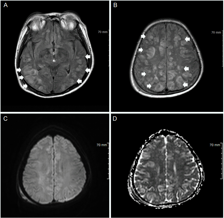
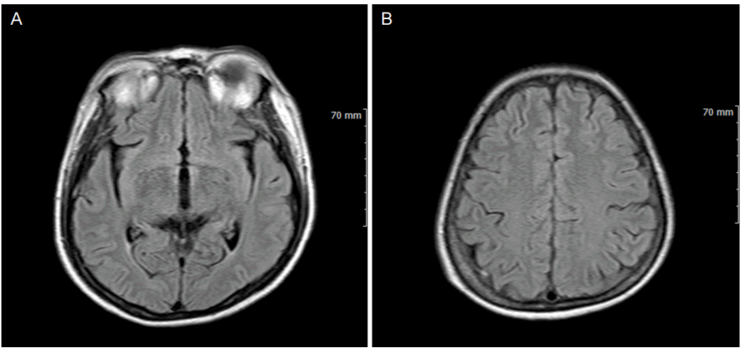
The patient developed seizures after admission. Intravenous lorazepam 4 mg was given for acute control of seizure, after which she underwent brain magnetic resonance imaging (MRI). Fluid attenuated inversion recovery demonstrated diffuse vasogenic edema in the bilateral cortical and subcortical white matters involving the frontal lobes. While diffusion-weighted imaging revealed nonsignificant high signals, the apparent diffusion coefficient map demonstrated high signal intensities in these regions (Fig. 1A-D). There was no history of cytotoxic immunosuppressive therapy. Perfusion MRI (MRP) showed no hyperor hypoperfusion at blood pressure levels of 140/50 mmHg (Fig. 2A-D). After close monitoring of blood pressure, her symptoms were resolved. Considering low recurrence rate of seizure, we did not prescribe any antiepileptic drugs. The 3 weeks follow-up MRI demonstrated complete resolution of previous lesions (Fig. 3A-B). This might be interpreted as atypical PRES.
DISCUSSION
PRES is a neurologic syndrome, diagnosed by clinical and radiologic features. The typical presentation includes headache, mental change, visual symptoms, and seizures. The characteristic MRI findings are consistent with vasogenic edema predominantly localized to the posterior hemisphere. It is not entirely elucidated why PRES favors the posterior circulation, but this might be related to a relative lack of sympathetic innervation at the level of the arterioles supplied by the vertebrobasilar system, compared with the anterior circulation [4]. However, PRES is not a posterior regional phenomenon. Additionally, more than 70% of patients with PRES are hypertensive, though significant proportions have normal or only mildly raised blood pressure [5]. Previous reports have demonstrated that PRES can occur in cases of atypical distributions, and features of imaging findings and normotensive settings [4,6]. The exact pathophysiology of PRES remains controversial. One currently favored theory is hypertension with autoregulatory failure. When the upper limits of autoregulation are exceeded, there is a development of blood vessel alteration, capillary bed injury, vasogenic edema, and hyperperfusion. However, it does not explain why PRES occurs in the absence of hypertension [5]. Furthermore, although this theory suggests cerebral hyperperfusion, some evidences demonstrate cerebral hypoperfusion [7]. Another earlier theory is associated with a systemic inflammatory process including immune system activation, endothelial injury, vasoconstriction and hypoperfusion. However, it is not clear why some cases of PRES occur without any inflammatory state [5]. Despite that, one large series of cases demonstrated that atypical distribution of PRES have a higher incidence than commonly perceived. PRES is not an entirely posterior phenomenon, but rather shows in a gradient-like pattern, reflecting the gradient of sympathetic innervations [4].
In our case, vasogenic edema occurred at relatively not very high blood pressure, and the imaging findings of MRI showed atypical appearances. Based on a previous report, although peak systolic blood pressure is usually between 170-190 mmHg, 10-30% of patients have normal or mildly elevated blood pressure. In clinical settings, uremia in CKD patients may mediate their effect on the vascular endothelium or other regions of vasogenic control. Furthermore, it is important to note that PRES is a dynamic process. The MRP in PRES is significantly increased in the acute hypertensive setting, but after the blood pressure has reduced, perfusion also tends to decrease [8]. Because contrast perfusion method was contraindicated in CKD patients, the exact perfusion mechanisms in these patients were not elucidated. However, because the MRI (including perfusion) was performed on this patient to rule out hyperacute ischemic stroke, this case might propose some critical information for the hemodynamic mechanisms of atypical PRES.
In conclusion, we suggest that MRP should be considered in the appropriate temporal framework, to avoid misinterpretation of other diseases, such as acute cerebral ischemia, cerebral venous thrombosis, encephalitis, and brain tumor, especially in patients with CKD.


