A case of Brown-Sequard syndrome caused by spinal cord infarction
Article information

Typical imaging hallmarks of spinal cord infarction include bilateral anterior or central cord lesions. However, unilateral hemicord lesions were rarely reported [1]. A 94-year-old woman visited our institution due to sudden right hemiplegia. Neurological examination findings were consistent with Brown-Sequard syndrome. The motor functions of the right arm and leg were given Medical Research Council scale grades of 1 and 2, respectively. Touch sensation was decreased in the right arm, trunk, and leg. Pain and temperature sensations were decreased on the left side.
Magnetic resonance imaging showed diffusion restriction and T2-hyperintensity of the spinal cord between C2 and C5 level (Fig. 1). Cerebral angiography revealed a right vertebral artery occlusion (Fig. 2). The ischemia of separated anterior spinal arteries or sulco-commissural arteries can provoke partial Brown-Sequard syndrome [2]. However, under the above conditions, posterior spinal artery territories remain spared.

Magnetic resonance imaging scans of cervical spinal cord obtained on admission date. (A, B) Diffusion-weighted imaging demonstrates diffusion restriction of the spinal cord between C2 and C5 level. (C) Sagittal T2-weighted image demonstrates high T2 signal intensity of the spinal cord between C2 and C5 level. (D) Axial T2-weighted image shows high T2 signal intensity of the right half of the spinal cord at C4 level.
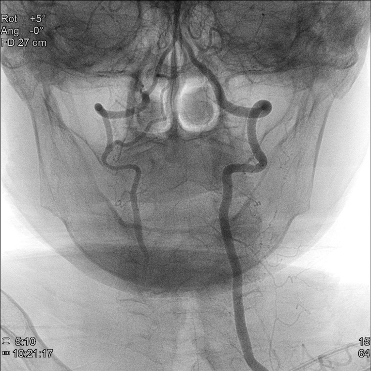
Percutaneous cerebral angiography of posterior circulation showing the occlusion of right vertebral artery from its origin. The left vertebral artery, on the other hand, is intact.
The radiculomedullary arteries are branches of radicular arteries arising from the vertebral artery. They form a Y-shaped branch in the cervical region supplying both anterior and posterior spinal artery territories [3,4]. Ischemia of the unilateral radiculomedullary artery could provoke hemicord infarction [4]. In our case, occlusion of the right vertebral artery might have caused an ischemia of the ipsilateral radiculomedullary artery.
Notes
Ethics statement
Ethical approval for this study was not needed in accordance with our Institutional Ethics Policy. This case report did not include any protected health information. Written informed consent was obtained from the patient.
Conflict of interest
No potential conflict of interest relevant to this article.
Author contributions
Conceptualization: SYS. Data curation: KL. Formal analysis: SYK. Project administration: KL, SYK. Visualization: SYK. Writing–original draft: SYK. Writing–review & editing: all authors.