| Home | E-Submission | Sitemap | Contact Us |  
Journal of Neurocritical Care > Epub ahead of print
Jeong, Yoo, Kim, and Kim: Basilar artery hypoplasia with persistent fetal circulations misinterpreted as basilar artery occlusion in a non-invasive angiography
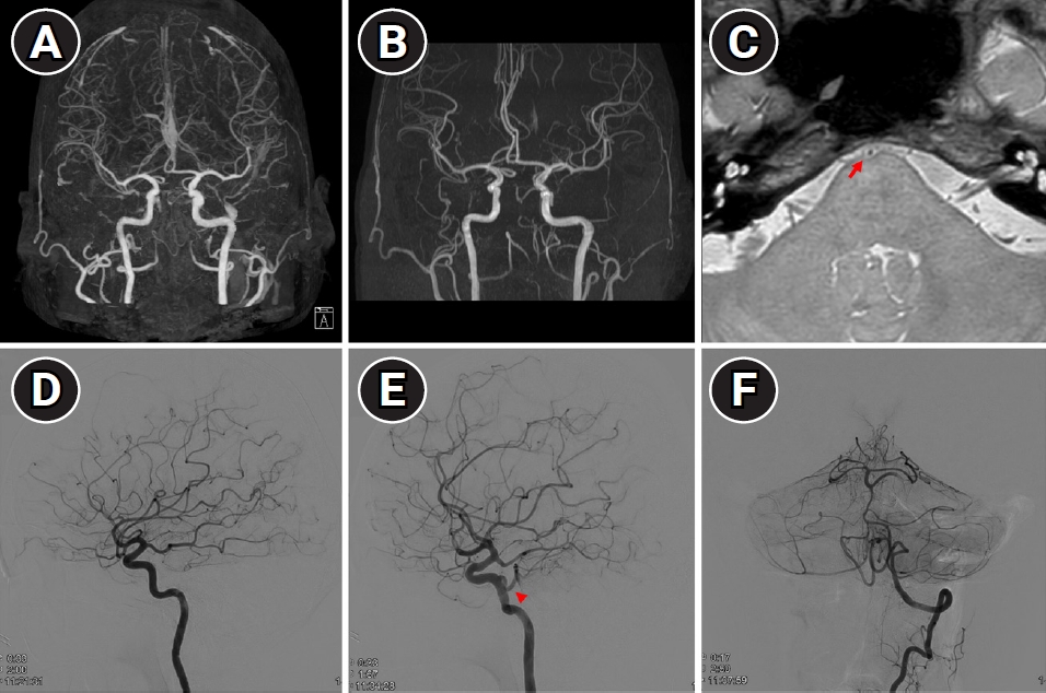
A 55-year-old woman presented with acute memory impairment, initially suspected as transient global amnesia. To rule out alternative causes, she underwent diffusion-weighted imaging (DWI) with time-of-flight magnetic resonance angiography (TOF-MRA) and computed tomography angiography (CTA). While the DWI showed no abnormalities, the basilar artery was not visualized in either CTA or TOF-MRA (Fig. 1A and B). Due to the non-visualized basilar artery, she was admitted for further investigation.
High-resolution magnetic resonance imaging (Fig. 1C) and conventional angiography (Fig. 1D-F) were subsequently performed. These examinations revealed a hypoplastic basilar artery with bilateral fetal posterior cerebral arteries (PCAs) and a left persistent primitive trigeminal artery (PPTA). Since basilar artery patency was confirmed, the patient was discharged without any interventions.
The incidence of PPTA is very low, with one Japanese cohort finding a mere 0.68% occurrence [1]. Among these cases, approximately 28% were presented with severe basilar artery hypoplasia. In our case, the presence of bilateral fetal PCAs further accentuated the hypoplastic basilar artery, creating an occlusion-like appearance. Notably, the proton density-weighted sequence in high-resolution magnetic resonance imaging helped differentiate occlusion from hypoplasia. Given the invasive nature of cerebral angiography, utilizing high-resolution magnetic resonance imaging in similar clinical situations could be a prudent alternative.

ARTICLE INFORMATION

Ethics statement
This study was reviewed and approved by the Institutional Review Board of Hanyang University Hospital (No. 2022-10-013). The need for informed consent from the patient was waived by the board.
Conflict of interest
No potential conflict of interest relevant to this article.
Author contributions
Conceptualization: YSK. Methodology: HYK. Formal analysis: HKY. Data curation: HKY. Visualization: SJ. Project administration: SJ. Writing–original draft: SJ. Writing–review & editing: SJ.

Fig. 1.
Multiple images of brain magnetic resonance imaging and conventional angiography. Computed tomography angiography (A) and time-of-flight magnetic resonance imaging (B) showed a non-visualized basilar artery. High-resolution magnetic resonance imaging (C) revealed hypoplastic basilar artery at the level of pons (red arrow). (D-F) Conventional angiography images demonstrate fetal-type posterior cerebral arteries and left persistent primitive trigeminal artery (red arrowhead).
jnc-230042f1.jpg

REFERENCES

1. O'uchi E, O'uchi T. Persistent primitive trigeminal arteries (PTA) and its variant (PTAV): analysis of 103 cases detected in 16,415 cases of MRA over 3 years. Neuroradiology 2010;52:1111-9.
crossref pmid pdf
Editorial Office
Department of Neurology and Critical Care, Seoul National University Hospital,
101, Daehak-ro, Jongno-gu, Seoul, 03080 Korea
TEL: +82-2-2072-0743 (10:00 AM ~ 4:00 PM) / +82-2-2072-1810   FAX: +82-2-3672-7553    E-mail: office@e-jnc.org

Copyright© Korean Neurocritical Care Society.                Developed in M2PI